Luxating Patella in Dogs.

Patella luxation, also known as a dislocated kneecap, is one of the most common orthopaedic conditions seen in dogs—especially small breeds, but also increasingly diagnosed in larger dogs. In some cases, patella luxation occurs alongside angular limb deformities, requiring advanced surgical techniques such as distal femoral osteotomy (DFO) for effective correction.
What Is Patella Luxation?
The patella (kneecap) normally sits in a groove at the end of the femur (thigh bone) and glides up and down as the knee flexes and extends. In dogs with patellar luxation, the kneecap slips out of this groove (luxates) - either medially (toward the inside of the leg) or laterally (toward the outside).
Patellar luxation may be congenital (present from birth) or acquired due to trauma. In many small breeds like Pomeranians, Chihuahuas, Yorkshire Terriers, and Poodles, the condition is often hereditary and linked to skeletal abnormalities, such as:
Shallow trochlear grooves
Abnormal limb alignment
Malformed femurs or tibias
In larger breeds (such as Labradors, Boxers and Akitas), patellar luxation is less common but tends to be more severe when it occurs, either due to injury or developmental issues.
Symptoms.
Intermittent skipping or limping in one hind leg
Sudden yelping or lifting of a hind limb while walking or running
Bunny-hopping gait, especially during running
Reluctance to jump or exercise
Stiffness or discomfort, especially after rest
Signs of discomfort when the knee is touched
In severe or chronic cases, untreated luxation can lead to osteoarthritis, muscle atrophy, or permanent limb deformities. It can also increase the risk of cruciate ligament injury disease or rupture.
Diagnosis.
Diagnosis is typically made through physical examination and observation of gait. Imaging such as X-rays or CT scans are often used to evaluate:
Severity of luxation
Shape and depth of the trochlear groove
Any associated angular deformities of the femur or tibia
Presence of osetoarthritis
Treatment Options.
Treatment depends on the severity of the luxation and the degree of symptoms that it is causing.
1. Non-Surgical Management (Mild or Asymptomatic Cases)
Weight control
Anti-inflammatory medications
Joint supplements
Physical therapy
2. Surgical Correction (Moderate to Severe Cases)
Surgical treatment is recommended for luxations which cause discomfort or affect mobility. Standard procedures include a combination of:
Trochleoplasty: Deepening the trochlear groove (the groove that the patella runs in)
Tibial Tuberosity Transposition (TTT): Re-aligns the patellar tendon via cutting the tibia bone and moving the section of bone that connects to the patellar tendon. The bone is fixated in position by 1 or 2 bone pins. Dr Davids performs this procedure using a newer technique (using a TTTT2 device) which has the benefit of earlier post-operative weight-bearing, faster recovery, and reduced post-operative pin-related complications.
Soft tissue reconstruction: Adjusts tension on surrounding tissues.
3. Distal Femoral Osteotomy (DFO): Advanced Surgery for Angular Limb Deformities
In some dogs, patella luxation is not just due to kneecap instability, but also due to abnormal angulation or torsion of the femur. This is particularly common in:
Large or giant breeds
Dogs with congenital limb deformities
Recurrent or failed patella surgeries
In these cases, standard surgical techniques are not enough to address the bony deformities causing the patella luxation. This is where Distal Femoral Osteotomy (DFO) comes in.
What Is DFO?
Distal Femoral Osteotomy is a corrective bone surgery in which a precise cut is made in the distal femur (near the knee), and the bone is realigned to correct:
Valgus deformity (limb angling outward)
Varus deformity (limb angling inward)
Rotational misalignment
Once corrected, the bone is stabilized with a bone plate and screws. This realignment allows the patella to track correctly within the trochlear groove, resolving the underlying biomechanical cause of luxation.
The benefits of DFO are:
Corrects the root cause of luxation due to skeletal deformity
Reduces stress on the joint, improving long-term mobility
Can be combined with TTT or trochleoplasty for full stabilization
Recovery typically includes:
8-10 weeks of restricted activity
Regular rechecks and X-rays to monitor bone healing
Gradual return to exercise with physical rehabilitation
Most dogs show excellent functional improvement after DFO when performed by an experienced orthopaedic surgeon.
Prognosis.
The long-term outlook for dogs with patella luxation are very good when appropriately treated. Dogs undergoing DFO for complex deformities also enjoy good outcomes, especially when surgery is done early and post-operative care is followed closely.
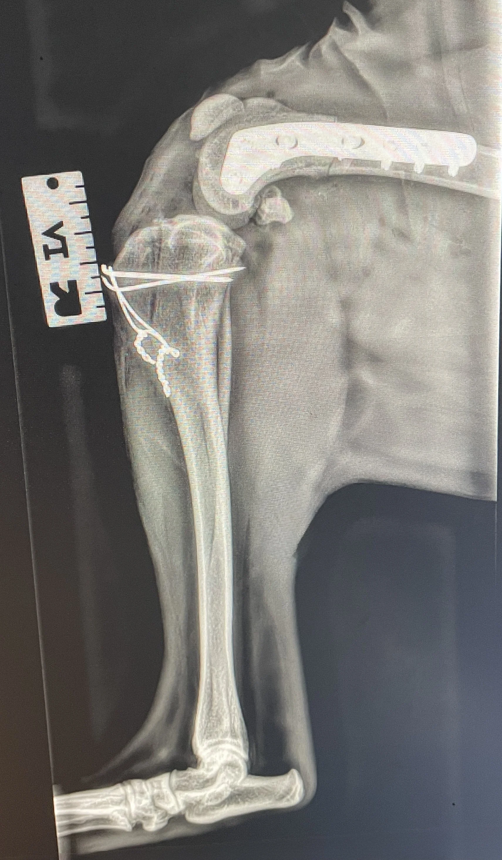
Post-operative radiograph of a DFO performed by Dr Davids.